Events

Optica End-User Workshop at Biophotonics Congress
25 April 2023
Hyatt Regency Vancouver, 34th Floor, Cypress Room, Vancouver, Canada
Focused for Industry. Free to attend
The End-User Workshop at the Biophotonics Congress is designed to bring together end-users from the biophotonics industry to exchange ideas on the latest developments in the field. Biophotonics is an interdisciplinary field that combines photonics, biology, and medicine to develop innovative solutions for healthcare, diagnosis, esthetics, and monitoring. These technologies are non-invasive, with high sensitivity, versatile, cost-effective, and require minimal sample preparation and at the same time with a big market in consumables. The workshop aims to provide a platform for end-users to share their experiences and discuss challenges in sensitivity, selectivity, photodamage, integration, and analysis. Main applications on microscopy, imaging, analytical instruments, therapeutic devices, sensing, optogenetic, and diagnostics will be addressed by end-users looking for collaboration with the industry. Participants will have the opportunity to engage, network, and collaborate to advance the development and adoption of biophotonics technologies in the industry.
Focused for Industry. Free to attend. Registration / RSVP required.
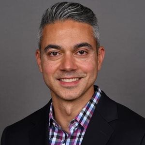
 Moderator - Helena Diez
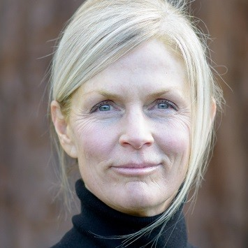 Moderator - Pamela Robertson
Agenda
3:00 PM: Doors open. Grab a coffee (or tea) and network.
3:20 PM: Invited speaker program. Bring your questions to this interactive workshop.
6:00 PM: Reception. Enjoy some refreshments, meet speakers one-on-one, network, connect.
Speakers
Speakers
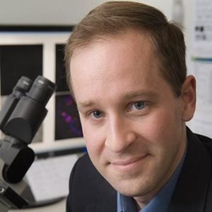
Conor Evans
Wellman Center for Photomedicine. Key interest: ultrasensitive oxygen detection and imaging technologies
Dr. Evans lab’s research is focused on the development and clinical translation of optical microscopy and spectroscopy tools, with specific interests in ultrasensitive detection of molecular markers, label-free imaging of drugs and tissues, and the imaging and quantification of tissue oxygenation. Dr. Evans has led the use of coherent Raman imaging technologies in biomedicine, and was the first to apply this imaging toolkit for the real-time visualization of lipids in skin in vivo. He has developed a number of imaging devices and methods in the fields of coherent Raman imaging, nanoscience, and “smart” wearable sensing technologies. A recipient of the NIH Director’s New Innovator Award, his efforts in the synthesis of bright oxygen sensors has resulted in the creation of new oxygen imaging technologies currently in clinical trials. He is a Royce Fellow of Brown University and has been honored with several awards, including the Goldwater Scholarship, NASA Space Grants, the National Science Foundation Graduate Fellowship, and the ASP New Investigator Award.
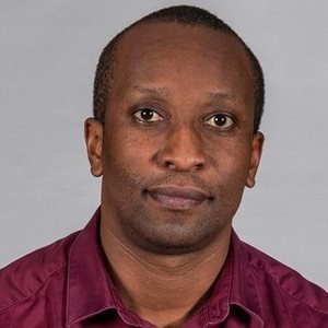
Raoul Vyumvuhore
SILAB. Key research: applications of Raman spectroscopy in the field of skin biophysics for better skin care
Dr. Raoul Vyumvuhore is a scientific researcher who has made significant contributions to the field of skin biophysics. He graduated as a pharmacist in St. Petersburg. After completing his degree, he moved to France to continue his education and pursue a career in scientific research. In France, Dr. Vyumvuhore obtained his Ph.D. in analytical chemistry at the University of Paris Sud. During his Ph.D. studies, Dr. Vyumvuhore's research has primarily focused on the application of Raman spectroscopy in skin research. After completing his Ph.D. studies, Dr. Vyumvuhore joined SILAB company, where he now manages the biophysics laboratory. In this role, he oversees the development and implementation of new techniques for the evaluation of natural cosmetic ingredients. He also leads the development of Raman spectroscopy-based methods for the characterization of skin and hair. In addition to optic’s research field, Dr. Vyumvuhore has also been involved in investigating mechanical properties of innovative cosmetic materials. Dr. Vyumvuhore has published several research articles in peer-reviewed journals and has presented his work at various international conferences. Overall, Dr. Vyumvuhore's work has helped to advance our understanding of skin structure and function and has the potential to lead to new diagnostic and therapeutic approaches for skin diseases, as well as the development of safer and more effective cosmetic products.
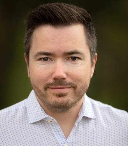
Josselin Breugnot
SILAB: key interest is the sequencing of skin microbiota genes or the classification of LC-OCT images in vivo.
Dr. Josselin Breugnot has been a data scientist and head of the data science unit for 15 years in SILAB's R&D laboratory. With a great attraction for the creation and scientific processing of images, he graduated from the French school Telecom Saint Etienne in 2007, member of the University of Lyon with a specialty in digital imaging. After a research master's degree in optics, vision and signal, he prepared a thesis on a perceptually justified color image processing framework based on a model close to human vision. He completed his doctorate in 2011 and has since published peer-reviewed articles on various topics such as the morphological study of mitochondria acquired by confocal microscopy, the sequencing of skin microbiota genes or the classification of LC-OCT images in vivo. using deep learning methods. At Silab, he ensures the optimization of the creation, extraction and quantification of data as well as the proper use of statistics for data analysis and during the statistical decision-making process. For this, it produces dedicated data processing tools in different programming languages, and thanks to a particular appetite for deep learning methods, deploys this revolutionary chapter of AI in the study of Silab dermo-cosmetic active ingredients.
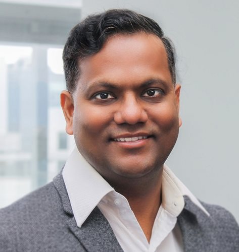
Sudip Shekhar
University of British Columbia: key interest is silicon photonics based biosensors for point-of-care and point-of-use applications.
Sudip Shekhar is an Associate Professor in the Department of Electrical and Computer Engineering at UBC. Dr. Shekhar received his B.Tech. from the Indian Institute of Technology, Kharagpur, in 2003 and the MSc and PhD degrees from the University of Washington, in 2005 and 2008, respectively. From 2008 to 2013, he worked as a Research Scientist in the Circuits Research Laboratory at Intel Corporation, Hillsboro, Oregon. He then joined the ECE department in 2013. Dr. Shekhar is a recipient of the IEEE Solid-State Circuits Society (SSCS) Predoctoral Fellowship, the Intel Foundation Ph.D. Fellowship, the Analog Devices Outstanding Student Designer Award, the Young Alumni Achiever Award by IIT Kharagpur, the IEEE Transactions on Circuit and Systems Darlington Best Paper Award and a co-recipient of IEEE Radio-Frequency IC Symposium Best Student Paper Award. He serves on the technical program committee of IEEE International Solid-State Circuits Conference (ISSCC), Custom Integrated Circuits Conference (CICC) and Optical Interconnects (OI) Conference. His research interests include circuits for high-speed interfaces, silicon photonics, radio-frequency transceivers and sensor interfaces.
Ali Azhdarinia
Univ. of Texas Medical Center, Key interest: real-time detection of neuroendocrine tumors during surgery
I am an Associate Professor in the Center for Translational Cancer Research at the University of Texas Health Sciences Center at Houston (UTHealth). My lab specializes in the development of bioconjugates for tumor-targeted delivery of radionuclides, fluorophores, and cytotoxic payloads. My primary research focus is in the development of a fluorescent somatostatin analog that enables real-time detection of neuroendocrine tumors during surgery. Our approach builds on the effectiveness of somatostatin analogs as validated tumor-targeting compounds and uses novel linker technologies to maintain their outstanding targeting and pharmacokinetic properties. The linkers uniquely allow us to minimally alter the chemical footprint of the parent compound while incorporating radioactive labels for cross-validation of fluorescence readouts. Accordingly, we utilize a variety of imaging techniques at the macro, meso, and microscopic levels for comprehensive agent evaluation.
